- Hormones
- Melatonin: Increases likelihood of sleep
- Glucocorticoids: Biomarker of circadian rhythm
- Growth Hormone: Accelerates growth in children
- Neurotransmitters
- Histamine: Increases alertness
- Norepinephrine: Maintains muscle tone
- Serotonin: Stabilizes wakefulness
- Brain Regions
- Basal Forebrain: Sleep + wakefulness
- RAS: Regulates sleep-wake cycle
- Thalamus: Sensory relay station
- Hypothalamus: Contains biological clock
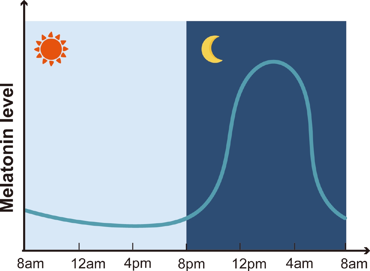
Different hormones, neurotransmitters, and brain regions work together to regulate sleep. Let's first learn about hormones–more specifically, melatonin, glucocorticoids, and growth hormone. Melatonin is a hormone synthesized from the tryptophan through a series of enzymatic reactions and mainly secreted by the pineal gland. Its synthesis is light-dependent as light information is processed to regulate its secretion (refer to previous section for more info). Melatonin performs several physiological functions, including the activation of MT1/MT2 receptors. By activating the MT1/MT2 receptors, melatonin inhibits SCN neuronal activity and lowers core body temperature, ultimately increasing sleep-propensity (likelihood of falling asleep at any given moment). Melatonin also helps regulate the circadian rhythm, contributing to sleep regulation.
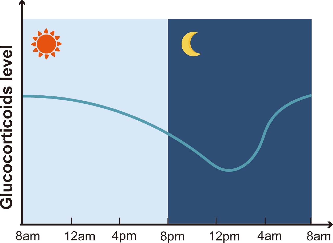
Glucocorticoids (Note: cortisol is a type of glucocorticoids) are hormones synthesized from cholesterol in the adrenal cortex. It performs important regulatory functions such as catalyzing catabolism of macromolecules (like proteins and lipids) and maintaining glucose homeostasis. They are reliable biomarkers of circadian homeostasis as they are characterized by a distinct circadian pattern, in which they peak in the early morning and gradually decline by midnight. As a result, dysregulation of this pattern indicates a circadian disruption. For example, total sleep deprivation and chronic circadian misalignment change cortisol levels of individuals. This results in changes in individuals' cortisol levels that are associated with increased risk of metabolic syndrome, musculoskeletal disorders, and neuropsychiatric disorders.
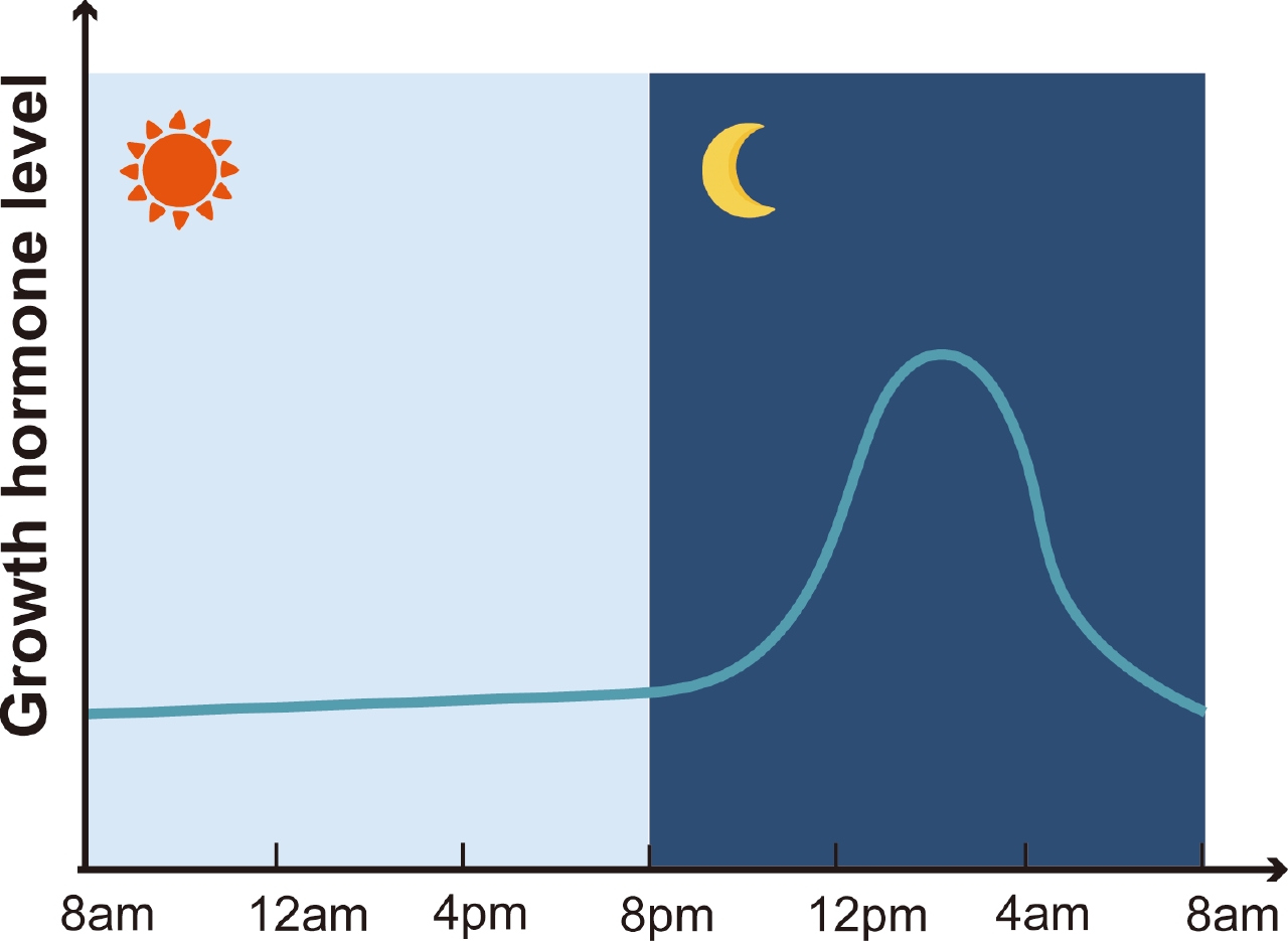
Growth hormone is a hormone that accelerates growth in children with hypopituitarism. It is synthesized in the anterior pituitary and plays many important roles in the body: promoting protein synthesis, maintaining muscle mass and strength, and accelerating wound healing. This hormone peaks during puberty, then continuously declines as individuals attain adult height. Its release is modulated by hypothalamic signals, including output from the SCN. It has a distinct circadian rhythm, where a pulsatile release occurs shortly after sleep onset in temporal association with the first SWS episode.
Now we will learn about involved neurotransmitters–in particular, histamine, norepinephrine, and serotonin. Histaminergic cells, located in the posterior hypothalamus of the brain, play a major role in promoting wakefulness. Active histaminergic neurons increase alertness while helping maintain sustained wakefulness. By contrast, GABAergic neurons promote sleep and significantly and directly inhibit hisaminergic cells and wake-promoting cells. Overall, the reduction in histamine activity leads to sleepiness and sleep onset, and sleep is only possible in the absence of histamine. When the posterior hypothalamus containing the histamine (wake center) is damaged, individuals suffer due to constant sleepiness. When the anterior hypothalamus and basal forebrain that contain GABA (sleep center) is damaged, individuals experience persistent insomnia.
Norepinephrine-producing neurons are active during wakefulness and inactive during REM sleep. Their inactivity during REM contributes to the characteristic features of REM sleep, atonia (temporary muscle paralysis). Norepinephrine is known to play a critical role in maintaining muscle tone during wakefulness, as observed by its absence during cataplexy, a sudden loss of muscle tone during consciousness. Similarly, their inactivity during REM sleep leads to muscle paralysis. Norepinephrine supports arousal indirectly by maintaining muscle tone and stabilizing wakefulness. Serotonin is a neurotransmitter that maintains arousal, regulates muscle tone, and manages some phasic events of REM sleep. It is inactive in sleep, especially in REM sleep, and its absence contributes to the distinct characteristics of REM sleep. The absence of serotonin removes suppression, causing PGO spikes (high voltage electrical activity) and release of eye movement and twitches, key indicators of REM sleep.
In the brain, the basal forebrain, reticular activating system, thalamus, and hypothalamus play instrumental roles in controlling sleep. The basal forebrain (BF) is a group of nuclei located at the base of the brain. It plays an important role in regulating cortical activity, which impacts wakefulness and certain sleep stages. The BF promotes cortical activity, especially during wakefulness and REM sleep. It contains a diverse population of neurons such as cholinergic neurons, GABAergic neurons (promote sleep), and glutamatergic neurons (support arousal). Such diversity allows the BF to be involved in both sleep and wake regulation and play an important role in sleep control.
The reticular activating system (RAS) is formed by a group of neurons extending diffusely from the medulla to the posterior hypothalamus. It is involved in maintaining wakefulness and receives afferent signals (sensory signals) from varied sensory systems (including light, sound, and movement). The RAS sends excitatory signals to the basal forebrain, thalamus, and hypothalamus, regions which activate the cerebral cortex and promote wakefulness. The thalamus plays an important role in modulating the sleep-wake cycle. It acts as a relay station for the glutamatergic sensory inputs to the cortex, meaning that all sensory signals pass through it before reaching the cortex. During wakefulness, thalamic neurons actively relay sensory information to the cortex, supporting alertness. However, during sleep, these neurons decrease their relay activity, resulting in the filtering or blocking of sensory input to the cortex. Overall, such reduction helps maintain sleep by preventing an overflow of sensory stimuli that may lead to awakening.
The hypothalamus is a region deep within the brain that manages the body's homeostasis and controls the sleep-wake cycle. The region contains the SCN which sets the pace of the circadian rhythm (refer to previous section). Particularly, the tuberomammillary nucleus (TMN), ventrolateral preoptic area (VLPO), and median preoptic area (MNPO) contribute to sleep regulation. TMN is a major wake center that promotes arousal through release of histamine. Turning off TMN histamine neurons makes one fall into deep SWS fast. Contrastingly, the preoptic area consisting of the VLPO and MNPO contain neurons that generate sleep. These neurons send inhibitory projections to areas that promote arousal, generating sleep. They are most active during NREM and REM sleep.